Loculated Pleural Effusion / 17 Best images about Disease of the lung on Pinterest ... / Complex septated, complex nonseptated, or homogeneously echogenic effusions are always exudates (fig.. Pocus demonstrated a large right sided loculated pleural effusion with associated septations and surrounding consolidation suggestive of a parapneumonic effusion. Pleural effusions in the intensive care setting. Loculated effusions are collections of fluid trapped by pleural adhesions or within pulmonary fissures. Complex septated, complex nonseptated, or homogeneously echogenic effusions are always exudates (fig. Pleural effusion is when fluid fills this gap and separates the lungs from the chest wall.
Pleural effusion is when fluid fills this gap and separates the lungs from the chest wall. This case highlights the atypical but unique presentation of a transudative pleural effusion and demonstrates the risk of repeated thoracocentesis complicating a simple clinical presentation. Complex septated, complex nonseptated, or homogeneously echogenic effusions are always exudates (fig. The largest pocket of fluid is present posteriorly at the right lung base, with associated atelectasis and minor consolidation. Pleural effusion is an accumulation of fluid in the pleural space that is classified as transudate or exudate according to its composition and underlying pathophysiology.
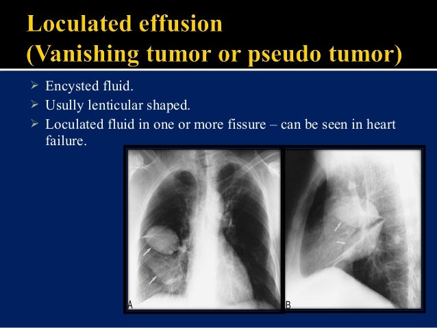
Loculated Pleural Effusion : State Of The Art Radiological ... from www.ccjm.org We present a unique case in which a patient presented to the ed in respiratory distress. Pleural effusions in the intensive care setting. Loculated malignant effusions however, are inherently resistant to the usual approaches because of nonexpanding underlying lung. Loculated effusions, defined as effusions that do not shift freely in the pleural space, occur when there are adhesions between the visceral and parietal pleura. Encysted pleural fluid is visualized between the right upper and middle lobe(s). A pleural effusion occurs when fluid fills this gap and separates the lungs from the chest wall. If the fluid cannot be drained, the lungs aren't able to expand and oxygenate the blood sufficiently. The largest pocket of fluid is present posteriorly at the right lung base, with associated atelectasis and minor consolidation.
Icu patients cannot sit up and the effusion layers posteriorly.
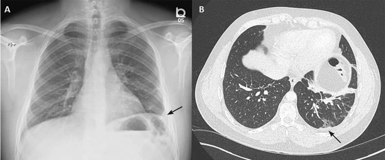
The largest pocket of fluid is present posteriorly at the right lung base, with associated atelectasis and minor consolidation. Loculated pleural effusion masquerading as mediastinal tumour had been reported but pleural effusion that conformed to the contour of a lung lobe is rare. We report a case in which loculated recurrent pleural effusion was treated by insertion of an indwelling tenckhoff catheter. Loculated effusions, defined as effusions that do not shift freely in the pleural space, occur when there are adhesions between the visceral and parietal pleura. In chf effusions are bilateral and more on right. Pleural effusion is an accumulation of fluid in the pleural space that is classified as transudate or exudate according to its composition and underlying pathophysiology. Most pleural effusions, whether free flowing or loculated, are hypoechoic with a sharp echogenic line that delineates the visceral pleura and lung. What are the different appearances of pleural effusion? Pleural effusion that is confined to one or more fixed pockets in the pleural space. Empyema is defined by purulent fluid collection in the pleural space, which is most commonly caused by pneumonia. Sometimes in the setting of pleuritis, loculation of fluid may occur within the fissures or between the pleural layers (visceral and parietal). 1 article features images from this case 21 public playlist include this case If the fluid cannot be drained, the lungs aren't able to expand and oxygenate the blood sufficiently.
If the fluid cannot be drained, the lungs aren't able to expand and oxygenate the blood sufficiently. Pleural effusions in the intensive care setting. Most effusions start like this and can be easily missed. Pleural fluid is seen extending to the right oblique fissure. This case highlights the atypical but unique presentation of a transudative pleural effusion and demonstrates the risk of repeated thoracocentesis complicating a simple clinical presentation.
Loculated Pleural Effusion Definition / Empyema Thoracis ... from image.slidesharecdn.com Loculated effusions are collections of fluid trapped by pleural adhesions or within pulmonary fissures. Loculated pleural effusion (427895005) recent clinical studies. Treatment may fail if the catheter is not placed optimally within the loculation or if the fluid is hemorrhagic or fibrinous. Most pleural effusions, whether free flowing or loculated, are hypoechoic with a sharp echogenic line that delineates the visceral pleura and lung. The largest pocket of fluid is present posteriorly at the right lung base, with associated atelectasis and minor consolidation. 681 views reviewed >2 years ago Most effusions start like this and can be easily missed. In chf effusions are bilateral and more on right.
A pleural effusion occurs when fluid fills this gap and separates the lungs from the chest wall.
Causes of an exudative effusion are malignancy, infection, or inflammatory disorders such as rheumatoid arthritis. We report a case in which loculated recurrent pleural effusion was treated by insertion of an indwelling tenckhoff catheter. Loculated pleural effusion masquerading as mediastinal tumour had been reported but pleural effusion that conformed to the contour of a lung lobe is rare. The largest pocket of fluid is present posteriorly at the right lung base, with associated atelectasis and minor consolidation. Loculated malignant effusions however, are inherently resistant to the usual approaches because of nonexpanding underlying lung. Icu patients cannot sit up and the effusion layers posteriorly. Loculated pleural effusion (427895005) recent clinical studies. In chf effusions are bilateral and more on right. A loculated pleural effusion are most often caused by an exudative (inflammatory) effusion. Ultrasonography is useful in cases of loculated pleural effusion for confirmation of the diagnosis and for marking a site for thoracocentesis. 681 views reviewed >2 years ago Loculated pleural effusion the pleura is a thin membrane between the lungs and chest wall that lubricates these surfaces and allows movement of the lungs while breathing. This type of effusion is empyema unless proven otherwise.
Ultrasonography is useful in cases of loculated pleural effusion for confirmation of the diagnosis and for marking a site for thoracocentesis. Pleural fluid is seen extending to the right oblique fissure. Loculated pleural effusion masquerading as mediastinal tumour had been reported but pleural effusion that conformed to the contour of a lung lobe is rare. Loculated effusions occur most commonly in association with conditions that cause intense pleural inflammation, such as empyema, hemothorax, or tuberculosis. Complex septated, complex nonseptated, or homogeneously echogenic effusions are always exudates (fig.
Parapneumonic Pleural Effusions and Empyema Thoracis ... from img.medscapestatic.com Initial testing … lupus pleuritis and other causes of pleural effusions in lupus patients. Loculated effusions, defined as effusions that do not shift freely in the pleural space, occur when there are adhesions between the visceral and parietal pleura. Pleural effusion is when fluid fills this gap and separates the lungs from the chest wall. In chf effusions are bilateral and more on right. Complex septated, complex nonseptated, or homogeneously echogenic effusions are always exudates (fig. Icu patients cannot sit up and the effusion layers posteriorly. If the fluid cannot be drained, the lungs aren't able to expand and oxygenate the blood sufficiently. Loculated pleural effusion the pleura is a thin membrane between the lungs and chest wall that lubricates these surfaces and allows movement of the lungs while breathing.
1 article features images from this case 21 public playlist include this case
Loculated effusions occur most commonly in association with conditions that cause intense pleural inflammation, such as empyema, hemothorax, or tuberculosis. In chf effusions are bilateral and more on right. 1 article features images from this case 21 public playlist include this case Causes of an exudative effusion are malignancy, infection, or inflammatory disorders such as rheumatoid arthritis. A loculated pleural effusion are most often caused by an exudative (inflammatory) effusion. Loculated pleural effusion (427895005) recent clinical studies. This type of effusion is empyema unless proven otherwise. This case highlights the atypical but unique presentation of a transudative pleural effusion and demonstrates the risk of repeated thoracocentesis complicating a simple clinical presentation. Encysted pleural fluid is visualized between the right upper and middle lobe(s). Complex septated, complex nonseptated, or homogeneously echogenic effusions are always exudates (fig. Pocus demonstrated a large right sided loculated pleural effusion with associated septations and surrounding consolidation suggestive of a parapneumonic effusion. A pleural effusion occurs when fluid fills this gap and separates the lungs from the chest wall. Pleural effusion that is confined to one or more fixed pockets in the pleural space.